Portal Vein Stenosis Following Liver Transplantation Hemodynamically Assessed with 4D-flow MRI before and after Portal Vein Stenting
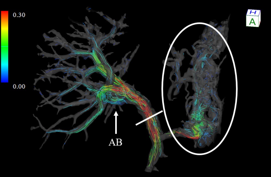
Anastomotic portal vein stenosis occurs as a postoperative complication of a living-donor liver transplantation, which leads to liver dysfunction. 4D-Flow MRI was performed on a 33-year-old woman who developed portal vein stenosis due to liver transplantation before and after stenting for portal vein stenosis. They have evaluated the flow pattern and flow volume of the portal vein and each branch. 4D-Flow MRI showed that the portal flow volume reduced and the heterogeneity flow distribution of portal branch blood flow volume before stenting. Improvements in these findings were observed after stenting.
This paper is the first case report to observe the hemodynamics of the portal system using 4D-Flow MRI. The hemodynamic evaluation of the portal system has previously been performed by angiography or portal angiography. 4D-flow MRI could be useful for non-invasively evaluating for understanding the hemodynamics, planning for treatments, and evaluating the outcome of the interventions.
Reference
Ryota Hyodo, Yasuo Takehara, Takashi Mizuno, Kazushige Ichikawa, Yasuhiro Ogura, and Shinji Naganawa, "Portal Vein Stenosis Following Liver Transplantation Hemodynamically Assessed with 4D-flow MRI before and after Portal Vein Stenting", Magnetic Resonance in Medical Sciences , Published Online (2020) 2020-0057.
Go To the Journal Page