Here, we present real-world examples published in academic papers, as well as YouTube videos shared with the subjects’ consent.
Videos published with the subject's consent
Case 1: Tetralogy of Fallot
Case 2: Tetralogy of Fallot
論文で公開された実例
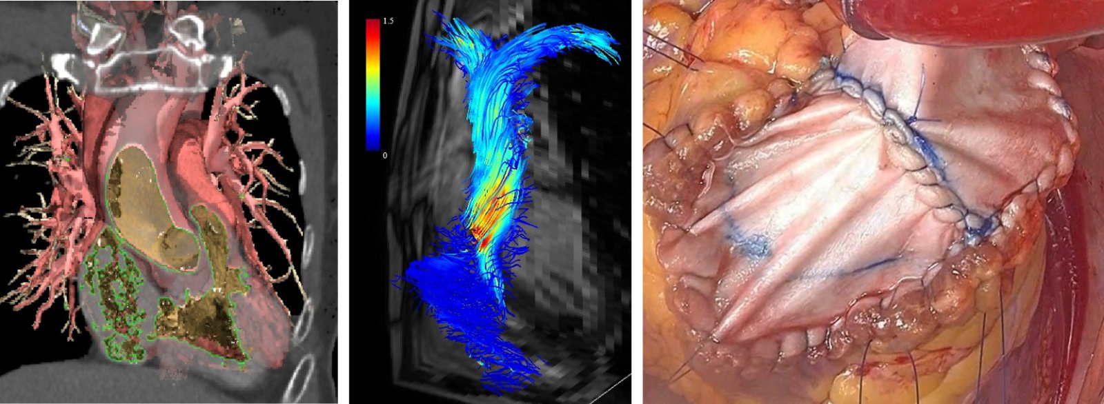
① A Case Report of the Correction of Tetralogy of Fallot That Had Remained Uncorrected Until Adulthood
Journal of Cardiothoracic Surgery. 2026;21(1):556 Read the paper
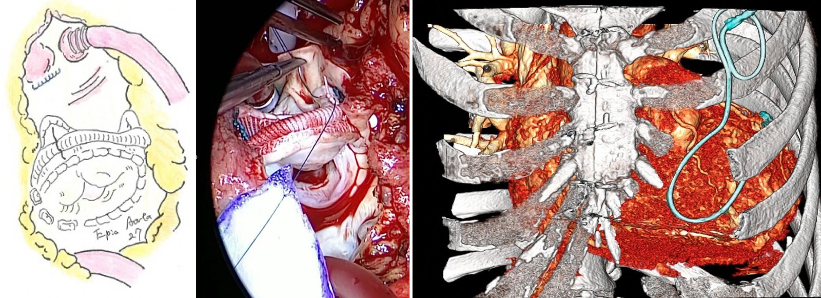
Clinical examples presented in the paper. Left: Narrow right ventricular outflow tract as seen in a preoperative CT simulation; Middle: Accelerated blood flow in the right ventricular outflow tract as seen in preoperative 4D flow MRI; Right: Surgery in which the pulmonary valve was replaced with a biological valve and the outflow tract was widened with a patch.
This is a case of Tetralogy of Fallot diagnosed following the onset of right heart failure in adulthood. This report details how Dr. Itaya made the diagnosis through blood flow analysis and performed intracardiac repair, including pulmonary valve replacement with a bioprosthetic valve.
In this condition, the right ventricular outflow tract is narrowed, leading to elevated right ventricular pressure and right ventricular distension. This can result in edema and shortness of breath, as well as potentially dangerous arrhythmias.
Even in patients who are told they have a hole in the heart (ventricular septal defect) but with low blood flow through it, relieving the narrowing at the outflow tract can lower right ventricular pressure and increase the volume of blood flowing through the defect.
With an accurate diagnosis based on blood flow analysis and appropriate repair, patients can lead a life free from worry.
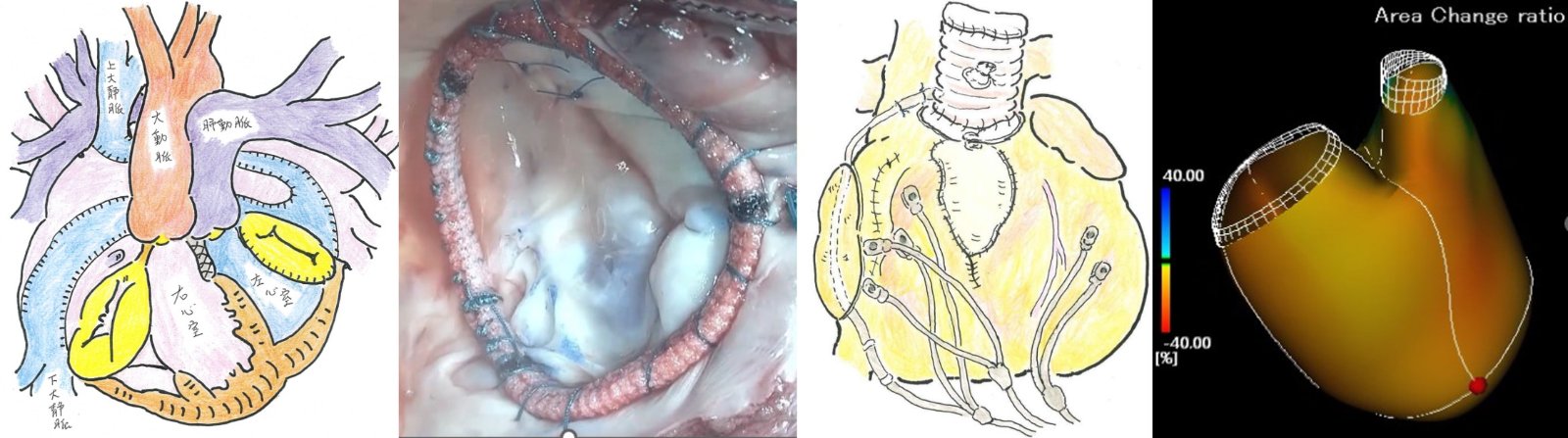
② A Case Report of Reintervention for Late-Stage Heart Failure in a Patient with Transposition of the Great Arteries and a Right Ventricular Ventricle
Asian Cardiovascular and Thoracic Annals. 2024;32(5):317-320 Read the paper
Case examples presented in the paper. From left to right: preoperative state of atrial blood flow reversal due to transposition of the great arteries; surgical creation of the systemic-ventricular tricuspid valve; an illustration of the surgery with a CRTD implanted; and postoperative right ventricular function.
In the past, there was a time when, in cases of transposition of the great arteries, blood flow was redirected within the atria, and the right ventricle was reconstructed to serve as the systemic ventricle (systemic ventricle). Even today, in cases of corrected transposition of the great arteries, the right ventricle may still function as the systemic ventricle.
Since the right ventricle is not exactly the same as the left ventricle, patients may develop heart failure, valvular disease (systemic ventricle atrioventricular valve and tricuspid regurgitation), or arrhythmias in adulthood.
Dr. Itaya treated a patient who was transported to the emergency room with ventricular arrhythmia triggered by a cold. To improve the patient’s cardiac function, he performed tricuspid valve repair and cardiac resynchronization therapy with defibrillation (CRTD), successfully restoring the patient’s heart so that they could once again enjoy traveling in good health.
The paper details specialized discussions on the management and techniques involved in reoperation, as well as how innovative 4D imaging was utilized.
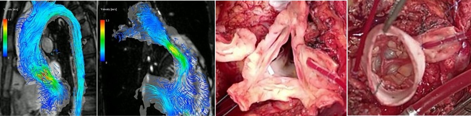
③ A Case Report of Double Root Replacement Surgery Involving the Aortic Root and Right Ventricular Outflow Tract Following a Ross Procedure
Journal of Surgical Case Reports. 2024;2024(5):rjae294 Read the paper
Case examples presented in the paper. Left: Preoperative 4D flow MRI of the left and right cardiac systems; Right: Aortic root replacement with native valve preservation.
Since there are no artificial valves that grow with the child, the Ross procedure—in which the pulmonary valve is excised and transplanted to replace the aortic valve—is sometimes performed for congenital aortic valve disease.
Because the pulmonary valve is not subjected to as high a pressure as the left ventricle, it can be replaced with an artificial blood vessel sewn to an artificial leaflet.
However, since the aortic valve after a Ross procedure was originally part of the pulmonary artery, sustained high blood pressure can cause the aortic root to bulge and form an aneurysm, leading to valve misalignment and regurgitation.
In addition, the artificial blood vessel with a pulmonary valve implanted during childhood may develop stenosis or regurgitation due to durability or sizing issues.
This is known as the “two-valve problem” following a Ross procedure and can become a serious issue in adulthood.
Dr. Itatani replaced the aortic root while preserving the patient’s own valve—which had been replaced during the Ross procedure—and simultaneously replaced the pulmonary valve with an adult-sized bioprosthetic valve inserted into a synthetic graft. Such surgery is a major procedure and a specialized treatment that requires accurate diagnosis and meticulous planning.
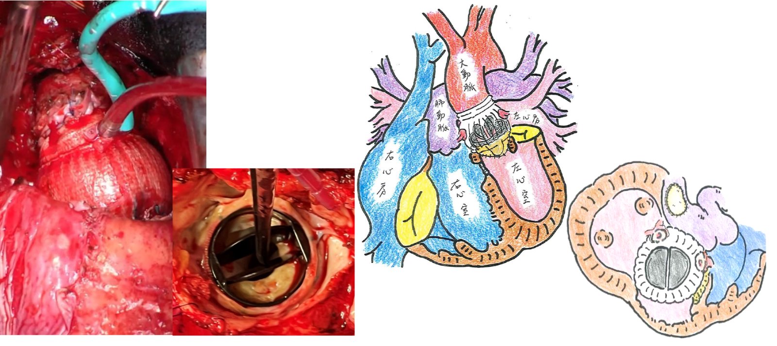
④ A Case Report of a Patient with Bilateral Great Vessel Right Ventricular Origin Syndrome Who Underwent Reoperation at the Base Due to Mechanical Valve Failure Following Mechanical Valve Replacement After a Great Vessel Switch Operation
Annals of Thoracic Surgery Short Report. 2024;3(2):464-468. Read the paper
論文で紹介された実例。左:手術所見とパンヌスのはった機械弁、右:解剖の概念図と手術シェーマ
In cases of congenital heart disease, complex anatomy may sometimes require multiple surgeries. This paper describes a case of posterior TGA—a form of double-outlet right ventricle—characterized by a displacement of the great vessels, in which a mechanical prosthetic valve was required following a Jatene aortic switch procedure performed during childhood. However, when that mechanical valve failed and the fragile aortic root began to dilate, Dr. Itatani conducted a detailed anatomical assessment. He performed aortic root replacement surgery (the Bentall procedure) to restore the anatomical right ventricular outflow tract, thereby preventing sudden death and enabling the patient to lead a safe life and return to work through further reoperation.
⑤ A case report describing pulmonary valve replacement and biventricular pacing from both the endocardial and epicardial sites (ventricular resynchronization therapy) for left and right heart failure in a patient with Tetralogy of Fallot
Journal of Cardiothoracic Surgery. 2024;19(1):98 Read the paper
Clinical examples presented in the paper. Left: Bioprosthetic pulmonary valve replacement; Right: Biventricular pacing using an epicardial lead
After repair of Tetralogy of Fallot, as the patient ages, not only does right ventricular failure due to pulmonary valve disease develop, but left ventricular function may also deteriorate.
Sometimes this is partly due to a loss of coordinated wall motion in the ventricles; in such cases, biventricular pacing may be performed to regulate ventricular contraction.
Although this can sometimes be anatomically challenging, Dr. Itatani combined biventricular pacing—using epicardial pacing for the left ventricle and transvenous pacing for the right ventricle—with pulmonary valve replacement using a biological prosthesis.
Reoperative procedures for congenital heart disease in adulthood not only improve life prognosis but also play a role in supporting patients’ ability to work and lead active lives.
⑥ A Case Report of Simultaneous Surgery for Left Atrioventricular Valve Regurgitation and Left Ventricular Outflow Tract Stenosis Following Repair of an Atrioventricular Septal Defect
Journal of Cardiothoracic Surgery. 2024;19:561 Read the paper
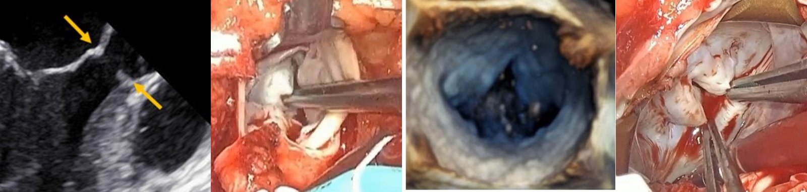
Case examples presented in the paper. Left: Echocardiographic findings and intraoperative findings of left ventricular outflow tract stenosis; Right: 3D echocardiographic findings and intraoperative findings of a left ventricular atrioventricular valve cleft
Atrioventricular septal defect is a common congenital heart defect. Although surgery is performed during childhood to close holes in the atrial and ventricular septa, these procedures often require simultaneous repair of the heart valves.
It is not uncommon for these heart valves to require reoperation later in life.
Additionally, due to anatomical characteristics, the portion of the aortic valve at the left ventricular outflow tract may become narrowed.
For patients with this condition, Dr. Itatani performed additional repairs on the atrioventricular valves based on the details of the childhood repairs, and also identified and surgically reduced the narrowing of the left ventricular outflow tract using CT imaging.
Congenital heart disease may require several surgeries throughout a patient’s life; therefore, it is essential to discuss treatment options while considering life plans and timing the procedures appropriately.
⑦ A Case Report on the Simultaneous Treatment of Pulmonary Regurgitation and Arrhythmia Following Correction of Tetralogy of Fallot
Journal of Medical Case Reports. 2019;13(1):30 Read the paper
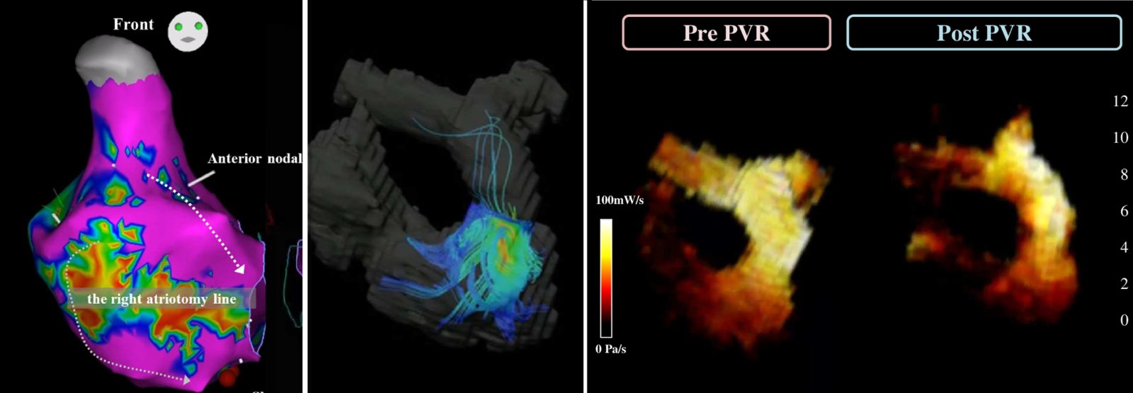
Case examples presented in the paper. Left: Atrial arrhythmia mapping; Middle: Pulmonary valve regurgitation as seen on 4D flow MRI; Right: Changes in energy loss before and after surgery
After repair of congenital heart disease, arrhythmias often occur due to right heart failure or scarring of the heart that developed during childhood.
Performing surgery on heart valves and blood vessels, as well as treating arrhythmias, within the context of congenital heart disease anatomy is an advanced and highly complex treatment that spans multiple specialties.
Dr. Itatani diagnosed pulmonary valve disease using MRI blood flow analysis, mapped the arrhythmia, performed a combined surgical procedure, and carried out the repair to enable the patient to lead a healthy life.
⑧ A Case Report on the Assessment of Right Heart Failure and Arrhythmia Following Ross Surgery Using Detailed Analysis of Cardiac Function, Blood Flow, and Electrophysiology
Journal of Medical Case Reports. 2020;14:113 Read the paper
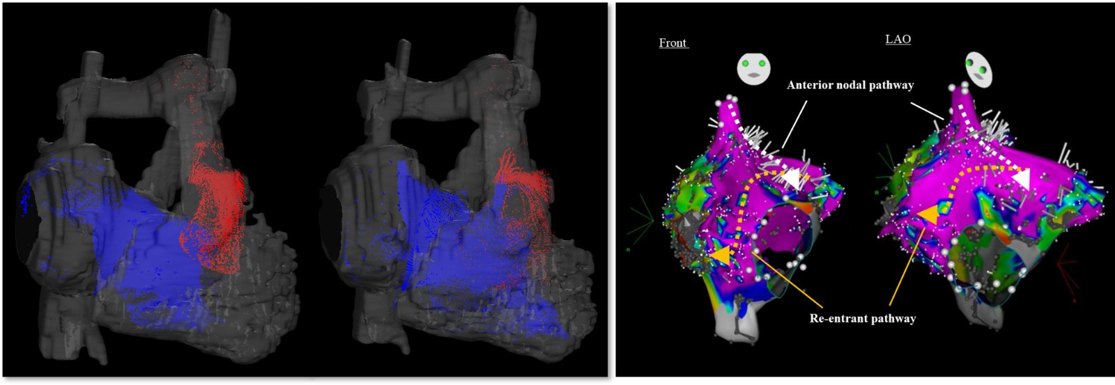
Case examples presented in the paper. Left: Pulmonary valve regurgitation as seen on 4D flow MRI; Right: Preoperative arrhythmia mapping
In cases of aortic valve disease in childhood, growth-related issues may arise; the Ross procedure—in which the pulmonary valve is excised and transplanted to replace the aortic valve—is sometimes performed, which can result in a “two-valve problem.”
Furthermore, right heart failure can sometimes trigger arrhythmias.
Dr. Itatani took a comprehensive approach to hemodynamics, heart failure, and arrhythmias, utilizing cutting-edge imaging to meticulously plan surgeries and treat these conditions simultaneously, working closely with patients to help them return to a healthy, active life.
In every developed country, this is a field where it is difficult to accumulate experience and data, where evidence is still insufficient, and where there are few precedents. That field is adult congenital heart disease.